Leica SP8 Confocal w/Two-Photon
Leica Leica SP8 Confocal w/Two-Photon (2025)
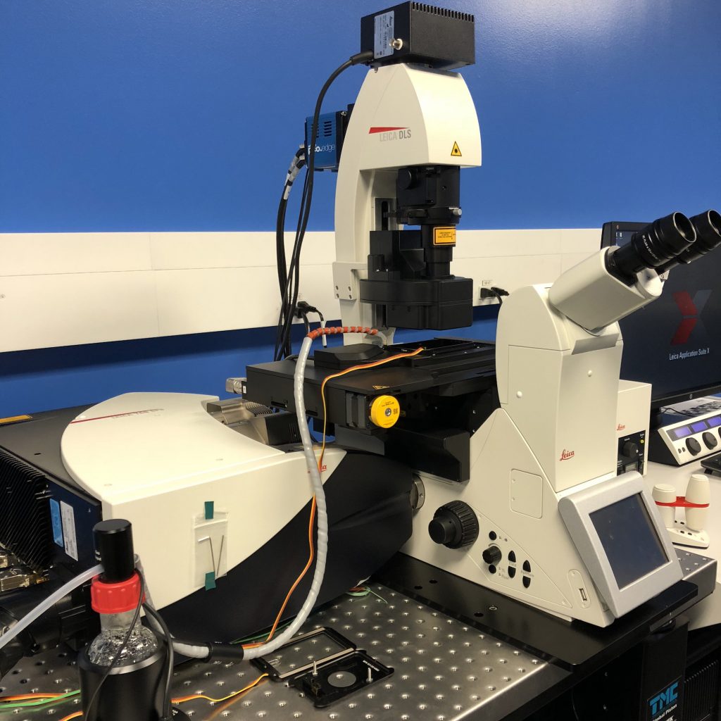
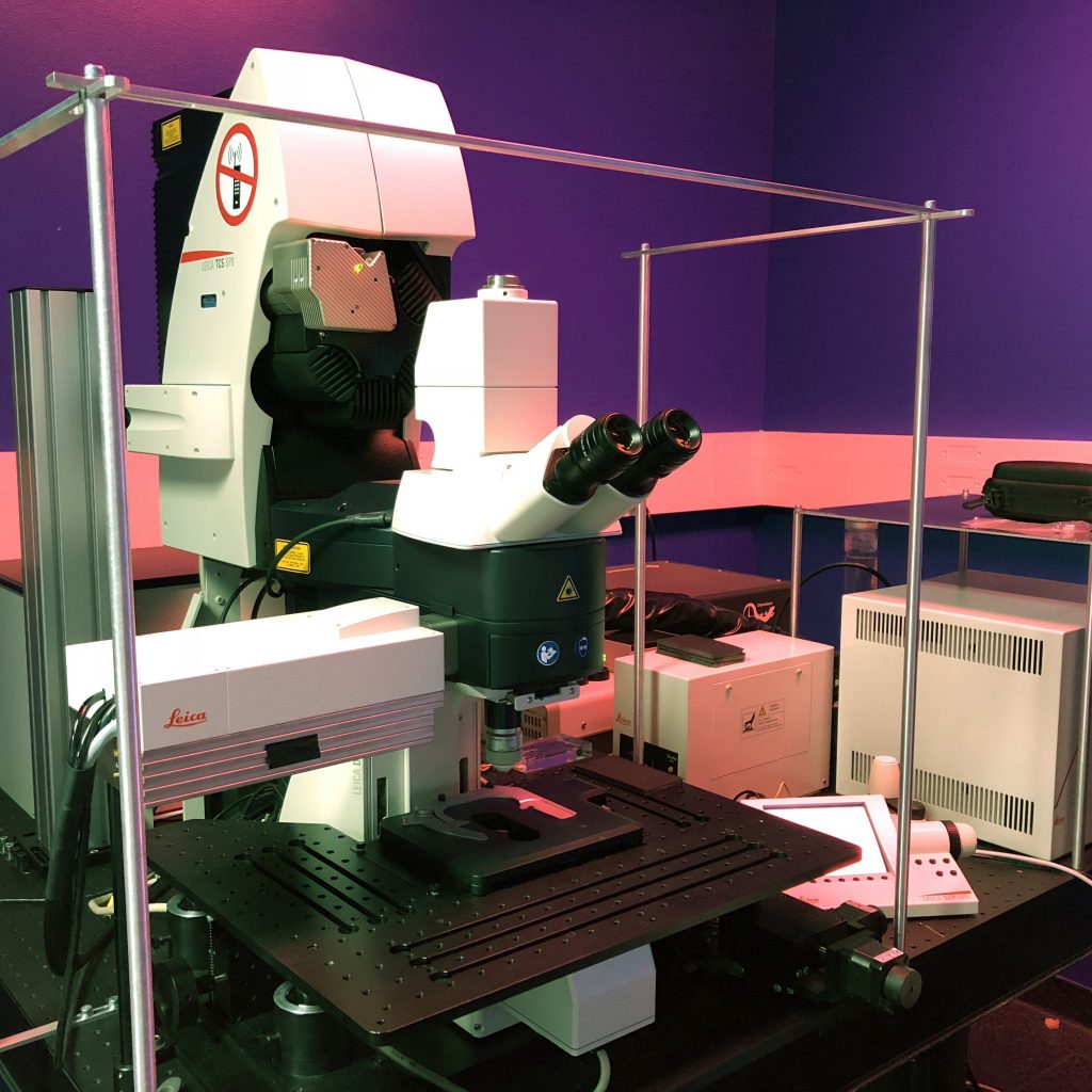
The Leica SP8 Confocal with Two-Photon is a versatile high-end microscope system that combines confocal imaging with multiphoton imaging, allowing for both high-resolution surface-level imaging and deep tissue visualization. The system is built on a modular Leica TCS SP8 platform and can be configured as an upright or inverted microscope. Leica SP8 single and two-photon confocal microscope – IPHYS ... Microscopes | SickKids Imaging Facility Key components and features Two-photon (multiphoton) microscopy This functionality is ideal for deep-tissue imaging because it uses lower-energy, longer-wavelength infrared light. The fluorescence excitation is limited to the exact focal point, preventing out-of-focus light from affecting the image and minimizing phototoxicity in thick samples. Deep tissue penetration: Longer wavelengths can penetrate significantly deeper into scattering tissues than visible light. Specialized detectors: The system uses highly sensitive, non-descanned hybrid detectors (HyD) for capturing signals deep inside the specimen. Infrared (IR) laser: Two-photon excitation is powered by a high-power femtosecond laser, often tunable within a range of 680 to 1300 nm. Confocal microscopy The SP8's confocal mode uses visible light to produce high-resolution, optically sectioned images of cells and tissues. This is especially useful for applications that require sharp contrast and precise localization of fluorescent signals. Tunable excitation: The white light laser (WLL) allows for the selection of any excitation wavelength between 470 nm and 670 nm for optimal fluorophore excitation. Spectral detection: A prism-based system and acoustic optical beam splitter (AOBS) enable the simultaneous capture of multiple channels and allow for spectral unmixing to separate overlapping fluorophores. Flexible scanning: The system offers both a standard galvanometer scanner for high-resolution imaging and an ultrafast resonant scanner for live-cell imaging. Applications The combined capabilities of the Leica SP8 with two-photon technology enable a wide range of advanced applications in life science research: Live-cell imaging: High-speed scanning and sensitive detectors enable dynamic imaging of living cells over time, and a built-in environmental chamber can control temperature and carbon dioxide. Deep tissue and in vivo imaging: The two-photon function is ideal for visualizing cellular processes in thick specimens, such as organoids, spheroids, or live model organisms. Fluorescence Lifetime Imaging Microscopy (FLIM): The system supports FLIM, which measures the decay rate of fluorescence to investigate molecular environments and interactions, such as Förster Resonance Energy Transfer (FRET). Super-resolution: Certain models can be equipped with STimulated Emission Depletion (STED) super-resolution to achieve resolutions of 50 nanometers or less. 3D image reconstruction: Software modules like LAS X allow for the acquisition and rendering of 3D images and tiling of large areas for complete sample visualization.
Capabilities
- ✓Confocal microscopy